![]()
|
|
|
Over the last 20 years have used a number of different model systems, both in vivo (whole organisms) and in vitro (cell culture systems). My favorite organisms are fish embryos both of the fathead minnow and more recently, the zebrafish. I enjoy working with fish embryos because they are easy to obtain and their embryos are fairly transparent so you can study development easily.
Even though my most of my work has been done with fish embryos, I seem to keep coming back to experiments involving the African Clawed Frog, Xenopus. Xenopus are great experimental animals because they have large eggs that are easy to manipulate and maintain and it is easy to do fertilizations manually.
I spent the summer in the laboratory of Michael Danilchik at Oregon Health Sciences University in Portland. The Danilchik Lab (http://www.ohsu.edu/som-pmcb/faculty/danilchik.html) is currently investigating a number of aspects of Xenopus embryogenesis including the activity of the cell membrane along the cleavage furrow during the first few cell divisions. My project involved injecting capped mRNA for farnesylated enhanced green fluorescent protein (EGFP-mem) into unfertilized Xenopus eggs, fertilizing the eggs and trying to study the activity of the membrane at the cleavage furrow with a confocal microscope. I first got used to working with the frogs and the video equipment in the lab. Below is a timelapse of normal cleavage in Xenopus (this is a large data file, don't try unless you have a fast connection).Normal Xenopus Early Cleavage (video 43Mb) We started by getting a the gene for EGFP-mem in a plasmid from a commercial source (Clontech). Clontech provides pEGFP-F vector that encodes for EGFP-mem. The pEGFP-F was then placed into pCS2+ vector for subsequent expression of mRNA. We used Ambion's commerically available mMessage mMachine large scale in vitro transcription kits for synthesis of capped mRNA's using the SP6 polymerase reaction.
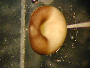
I then pulled some needles and tried injecting 10 nl of a maker dye as controls to see if normal development could occur. This was challenging, because when you poke a Xenopus egg, it will often activate the egg and cleavages will start. We tried to minimize this artificial egg activation by keeping the eggs cool and maintaining them in a low calcium solution. Unfortunately, the jelly layer surrounding the unfertilized eggs provided a formidable barrier to the needles I was using. As a result, lots of pressure was needed to get the needle in the egg and abnormal development resulted (see photo and timelapse movie below). An example of a bad injection experiment. (video 14Mb). In this video, normal embryos are on the right side and those that were injected are on the left side. Note that few of the injected embryos activate and develop.
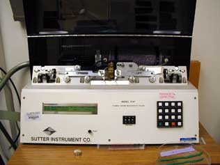
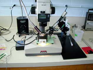
Trying to get through the jelly layer This was not working out, so I went back to the process of making the needles for injection, I tried lots of different kinds of micropipetts, and settings on the pipette puller. I eventually went to a wider filament which gave the nice looking tip, but I was still having problems with the injections. I found it was necessary to bevel the pipette tips to make it through the jelly layer.
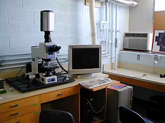
Micropipette Puller Needle Beveling Rig Once the protocol for injection of the capped EGFP-mem mRNA was worked out, we started to see if we would get expression of the protein. With the first series of experiments, expression would occur, but not until later cleavage stages. This was interesting for me because the expression of the green fluorescent protein allowed for the visualization of a number of membrane structures. Below is a confocal composite image of a late cleavage embryo and a highspeed timelapse confocal film taken on the Danilchik's lab new BioRad confocal microscope.
The new BioRad Confocal Microscope EGFP-mem-stained Xenopus embry
High-speed timelapse movie of EGFP-mem-stained cillary tuft in Xenopus larva (video)
It was very difficult to observe the activity of the membrane at the cleavage furrow for a number of reasons. First the cleavage furrow is a very dynamic site and it will not stay in the confocal field of focus for more than a few seconds. Second, the activity of membrane protrusions can not be visualized (at least not by me) real time. Rather, they can be seen only after taking a timelapse stack of photos assembled into a movie. What appeared was a series of membrane protrusions and microvilli that appear like "dancing worms" at the border of new and old membrane at the cleavage furrows. Below is a This is a confocal timelapse image of a 4-cell Xenopus embryo that has been injected with capped EGFP-mem. "Dancing Worms" of membrane in Xenopus cleavage furrow (video)
|