![]()
|
|
|
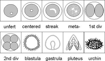
LAB 8 ECHINODERMS - FERTILIZATION AND EARLY SEA URCHIN DEVELOPMENT I. Introduction Our next model organism is the sea urchin which has been used extensively to study the events of fertilization and cleavage. The egg is small enough to be seen under the microscope, the gametes are produced in great abundance, and fertilization occurs naturally outside the body in sea water. The cytoplasm is relatively clear, so cleavage and gastrulation are easily observed. There are a number of objectives of this lab, they include: experience in the scientific method by designing your own experiments, observation of changes at fertilization of sea urchin eggs, artificially activation of eggs, investigation of the role of calcium in fertilization and probing for the existence of maternal RNA. The general timing of events surrounding fertilization in the sea urchin is as follows. The sperm become activated and undergo the acrosome reaction, shooting out a long filament. The sperm then penetrates the jelly coat that surrounds the egg, and the plasma membranes fuse. That moment of first contact sets off a series of ionic and metabolic events, some designed to block any other sperm from entering, some in preparation for developmental events leading to cleavage. The early block to polyspermy is a membrane ionic permeability event, while the late block to polyspermy results when cortical granules release their contents outside the cell to form the fertilization membrane, which lifts off and hardens. A. 0-60 seconds after fertilization 1. Small influx of Na+ 2. Release of intracellular Ca++ 3. Cortical granules fuse and release contents 4. Increase in oxygen utilization 5. Intracellular pH increases B. 5-90 minutes after fertilization 1. Increase in K+ conductance 2. Protein synthesis rate increases dramatically 3. Pronuclei fuse 4. First cleavage Cleavage in the sea urchin proceeds in a predictable fashion. The first two cleavages are meridional (longitudinal) and the third is equatorial, giving rise to eight equal-sized blastomeres. It is not until the forth cleavage that the smaller micromeres are formed at the vegetal pole. The sea urchin eggs have an animal pole and a vegetal pole that define the animal/vegetal axis. The animal hemisphere gives rise to epidermal structures and the vegetal pole generally gives rise to endodermal structures. The approximate time sequence of development is given below when we use the walk in cooler in the prep room. Developmental times vary with the incubation temperature:
1. Formation of fertilization envelope 2-5 minutes 2. First cleavage 1-2 hours 3. Second cleavage 1 hour later 4. Blastula 24 hours 5. Hatching 7-12 hours 6. Gastrula 48 hours 7. Pluteus Larvae 48+ hours
II. Procedure One objective of this exercise is to provide an opportunity to observe living gametes. Another is to give you an opportunity to design experiments to test some of the ideas used to explain mechanisms of fertilization. In designing an experiment, the most crucial (and often most difficult) aspect is to have a clear question in mind. Next decide how you will go about answering the question. These methods should be well thought out (in advance of the lab period) so as to help you decide between two possibilities or give an unequivocal answer. Next, the possible results should be anticipated and an explanation postulated even before the experiment is undertaken. Time spent in good planning means less time for repeating or scrapping a poorly planned experiment. You will now investigate the process of fertilization and early cleavage in sea urchins. Although you will be given access to live material and various chemicals, you will need to design your own experiments to answer some (four) questions. You will be working in groups of two. You should write up your results in your lab notebook in the format: a-question, b-method, c-result, and d-conclusion. Be sure you include controls in each experiment, and note what the controls were controlling for.
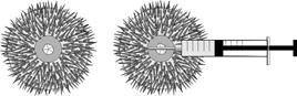
Problem 1 Can sperm fertilize an egg? How do we know the sperm are viable? Problem 2 Is there anything besides sperm that will cause the fertilization membrane to lift off? Hint: You know that Ca++ ions are involved and these ions among others are present in sea water. Problem 3 * Which of the structural proteins (microtubules or actin microfilaments) are involved in: a-the process of mitosis (karyokinesis), b-cell division (cytokinesis) in the early sea urchin embryo? Problem 4 * Is RNA production necessary for fertilization and early cleavage to occur? * Optional Below are the procedures for obtaining gametes, you must design your experiments using these procedures to answer the problems posed. Check the Materials section to see what is available for you to answer these questions. All Animals 1. Eggs and sperm of the sea urchin are collected by injecting 0.15 ml/inch of width of 0.55 M KCl with as small a needle as possible (25 gauge or so) needle into the body cavity at diametrically opposite points in the peristome (the soft integument surrounding the oral cavity). To avoid contamination of sperm and eggs, use a different syringe for each animal. Shake gently after injection to mix the KCl around, but be careful, as shaking too hard will kill the urchin. Then place the urchin with its oral side down on a chilled glass petri dish and look for gamete release. Do all work under clean conditions with cold, < 8oC water. When in doubt, chill all sea water and dishes that come in contact with gametes.
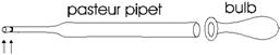
1. Within 5-15 minutes, males will produce a whitish mass of sperm suspension. Once the urchin has been determined to be male, wipe excess water from between the spines around the genital pore with a kimwipe. Then start collecting the sperm with a pasture pipette and place it is a chilled small test tube or beaker. "Dry sperm" may be stored for some time. When the sperm get into sea water, they have a very short live span. So it is important not to get sea water into the “dry” sperm stock.
2. Appropriate sperm dilution is essential for successful fertilization. Too low a sperm concentration results in fewer eggs being fertilized (safer than too high). Too high a sperm concentration will result in polyspermy (more than one sperm per egg) and abnormal development of the embryo. There are a number of built in mechanisms to help prevent polyspermy, but they are not full proof and at very high sperm concentrations they can be overwhelmed. Add "one notch" of sperm (2mm?) from a pasture pipette to 10 ml of sea water for fertilization under the microscopes and 2mm to 50-100 ml sea water for a normal development series. You may have to adjust this dilution up or down if necessary to get proper fertilization levels without polyspermy). Always use this quantity of sperm as the starting point for dilutions.
2 mm This is referred to as the STOCK SPERM SUSPENSION. A single drop of this suspension will be enough to fertilize 5 ml of a dilute egg suspension (1-2%). A single drop can be used to demo fertilization under a microscope. Although this is too many sperm for proper development, the embryos are unlikely to develop under a microscope for a variety of reasons (heat from lamp will make them too warm, the slide will dry out, etc.). This does, however, allow the student to witness, first hand, the raising of the fertilization membrane. Dry sperm that is added to sea water should be used within 10 minutes. Do not use to concentrated a sperm solution, if you do, you may get polyspermy which will kill the embryo. Sperm should be checked for motility under high-dry magnification. If they are not swimming, there is a good chance they are bad. 3. When sperm contact an egg they undergo the acrosome reaction in which the acrosome bursts open, releasing hydrolytic enzymes. A long filament forms that facilitates fusion of the sperm and egg plasma membranes. You can observe the acrosome reaction in vitro by placing sperm in some sea water where eggs have been sitting. Females 1. Female urchins are inverted with the oral side uppermost over the top of a small sterile egg cup or beaker filled with enough sea water to cover the genital pore. The ripe gametes will be shed for a period of 10-30 minutes, but you should start your experiments when you have enough eggs to work with. It has been estimated that a ripe urchin contains 1011 sperm or 8 X 10⁶ eggs. 2. Check the appearance of the eggs, they should be uniformly round, should have no “blebs”, if they have a large, clear nucleus they are probably immature, see below:
Immature Egg- No Good Mature Egg - size of Mature Nucleus 2. Gently wash the eggs 2-3 times after collecting the eggs by gently decanting the supernatant water and replacing it with fresh sea water. The final volume of sea water in the cup should be about 100 ml. This procedure removes broken spines, KCl and coelomic fluid, which inhibits the fertilization process. 3. Egg density should be no more than 1 cell layer thick on the bottom of the egg cup with 100 ml of sea water. Eggs can be stored at their normal temperature for up to 6 hours.
Normal Fertilization
2. Make up sperm stock solution as described above that will be viable for only about 10 minutes. Take a drop in a pasture pipette and place in under the coverslip with the eggs and observe.
________________ ______________(___________)____________
III. Questions 1. Describe the experimental design you used to solve problem #1 and #2. 2. Include detailed drawings (with labels) of: A. Fertilized and unfertilized eggs B. Embryos at 24 and 48 hours - you must come in to check on embryos 3. Discuss what may have caused problems with fertilization or later development, how could these problems be avoided.
VI. Materials 1. A23187 a calcium ionophore can cause the opening up of calcium channels in the plasma membrane. Dissolve 1 mg A23187 in 1 ml of 100% DMSO, then dilute 1:100 in sea water to give 10 ug/ml. 2. Actinomycin D blocks protein synthesis specifically by blocking the production of mRNA. Dissolve 2 mg in 50 ml sea water, then dilute 1:10 just before use to give 20 ug/ml. 3. Colchicine binds to tubulin, the major protein component of microtubules (which make up the mitotic spindle). Colchicine added to cells causes the microtubules to dissolve (has no effect of microfilaments). Dissolve 300 mg colchicine in 100 ml sea water. 4. Cytochalasin B acts on and dissolves microfilaments (has no effect on microtubules). Microfilaments are responsible for a number of activities in the cell including: phagocytosis, pinocytosis, exocytosis and cytokinesis. Dissolve 200 mg cytochalasin-B in 100 ml sea water. 5. About 5 liters of Sea Water on ice (28.32 gms NaCl, 0.77 gms KCl, 5.41 gms MgCl2@ 6H2O, 7.13 gms MgSO4@ 7H2O, 1.18 gms CaCl2, in 1000 ml distilled water, finally 0.20 gms NaHCO3 added after all others have dissolved) 6. 10-3 M EDTA Ca++/Mg++ Free Sea Water (30.80 gms NaCl, 0.77 gms KCl, 0.20 gms NaHCO3 in 1000 ml distilled water) 7. Gravid sea urchins (Woods Hole 508-548-3705 or Pacific Bio-Marine Labs 213-822-5757). The two most commonly used species are Panamanian urchin Lytechinus pictus, and the purple urchin, Strongylocentrotus purpuratus. 8. DMSO 9. 0.55 M KCl (50 ml) -Make up in e-pure water 10. 10, 20, 100 and 150 ml Beakers 11. Test tubes with racks and Watch Glasses 12. 5 ml, 10 ml and Pasture pipettes and bulbs 13. Syringes with small gauge (20 and 23 gauge or smaller) needles 14. Ice Buckets, modeling clay 15. Microscope and Depression Slides and Cover Slips 16. Dissecting Scopes With High Intensity Light Sources 17. Compound Microscopes and Hemocytometers 19. 10 and 25 ml graduated cylinders 20. Marking Pens 21. Transilluminator or high energy UV light source 22. Camera mounted on microscope to project fertilizations on screen
V. References 1. Giudice, G. 1973. Developmental Biology of the Sea Urchin Embryo. Academic Press, NY pp 63-86. 2. Steinhardt, R.A. and Epel, D. 1974. Activation of sea urchin eggs by calcium ionophore. Proc. Natl. Acad. Sci. USA. 71:1915-1919. 3. Biroc, S.L. 1986. Developmental Biology, A Laboratory Course With Readings. Macmillian Pub. Co. NY. 4. http://www.stanford.edu/group/Urchin/first.htm
|