![]()
|
|
|
LAB 3 ANATOMY OF THE ENDOCRINE SYSTEM
I am trying to mix up experimental labs with more traditional observational labs when possible. The observational labs are very useful to understand the structure, location and integration of endocrine structures. For this lab we will be doing a dissection of the fetal pig and the rat to locate the following endocrine organs:
Thyroid Adrenal Glands Pancreas Testis/Ovary Pituitary/Pineal
First get your dissection instruments and a dissection tray. Rinse the pig in the sink then make the following series of cuts to expose the organs in the thoracic and abdominal cavities. When you cut through the body wall with a pair of scissors, lift the body wall toward you so that you do not cut into any internal organs.
Once the body cavities have been exposed, again rinse out the inside of the pig. Then use string to tie the limbs around the bottom of the dissecting tray to allow the body cavity to stay open.
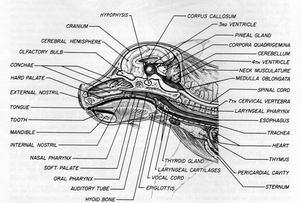
Identify the thyroid gland, it should be a dark solid organ on the ventral aspect of the trachea.
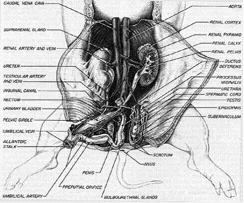
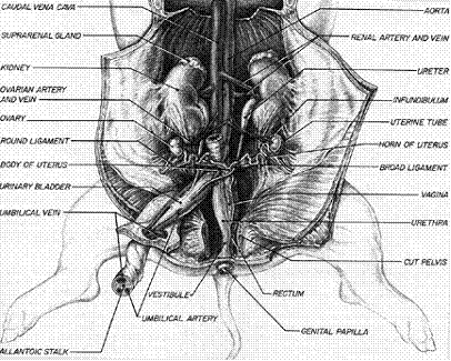
Next identify the pancreas, then the gonads in both a male and female pig.
Male Structures
Female Structures
If working with live rats, see appendix A on small mammal surgery. Once you are familiar with the procedures, place your rat on its back in a dissecting pan. In laboratory specimens there is usually an incision through the skin on the ventral surface of the neck for embalming and injection of the blood vessels with colored latex. Insert a blunt probe into this incision and loosen the skin from the underlying muscles of the body wall. Using your scissors, extend the incision posteriorly to a point just in front of the urogenital opening(s). Loosen the skin as you go and be careful not to cut the underlying tissues. Cut to the side of the urogenital and anal openings on either side and up around the base of the tail until the two incisions meet.
Return to the neck region and extend the mid-ventral incision anteriorly to the middle of the lower jaw. Next, make a lateral incision from the angle of the lower jaw upward to a point just behind the ear on each side. (Do not connect these incisions mid-dorsally.) Make a lateral incision from the ventral midline in the anterior thoracic (chest) region to the wrist along the medial surface of each forelimb. Similarly, make a lateral incision from the ventral midline just anterior to the urogenital opening(s) to the ankle along the medial surface of each hindlimb.
Throat region: A pair of large, dark submandibular salivary glands are located ventrally just behind the lower jaws. Do not confuse these glands with lymph nodes which are also located in this region and are smaller, rounded structures superficial to the salivary glands. A slightly smaller pair of parotid salivary glands are dorsal and lateral to the submandibular glands below the ear.
Neck region: locate the esophagus which transports food and fluids from the pharynx to the stomach in the abdominal cavity. It is found just behind and parallel to the trachea or windpipe. You will have to tease away some of the connective tissue surrounding the trachea and reflect it to one side to see the esophagus. Be careful not to remove the lobes of the thyroid gland, which are located on either side of the trachea just posterior to the larynx or voice box.
Opening the Abdominal and Thoracic Cavities: In order to see the remaining organs of the digestive system, the body cavities of the thorax and abdomen must be opened. With the rat lying on its back, make an initial midventral incision in the abdominal wall about an inch anterior to the genital region. The body wall is very thin at this point. Be careful not to cut too deeply. Lift the body wall away from the underlying organs and continue the midventral incision posteriorly to a point just anterior to the urogenital opening(s). Then continue the incision anteriorly toward the thoracic region.
At the anterior end of the abdominal cavity you will encounter the diaphragm, a sheet of muscle and connective tissue that separates the abdominal cavity internally from the thoracic cavity. Continue the incision through the ventral thoracic wall, veering slightly to the left of the midline to avoid cutting through the bony sternum (breast bone). The costal cartilages in this area are much easier to cut through that bone. When you reach the anterior end of the thoracic cavity move back to the midline at the base of the neck. You should now be able to trace the trachea from the neck region down into the thoracic cavity.
Make a pair of lateral incisions through the body wall immediately posterior to the diaphragm. Then carefully detach the diaphragm from the inner surface of the body wall. This will make it possible to spread open the rib cage to reveal the contents of the thoracic cavity without tearing the diaphragm. Finally make a pair of lateral incisions through the body wall at the posterior end of the abdominal cavity just in front of the urogenital region.
This will allow you to examine the organs of the abdominal cavity. Before continuing you may need to rinse out them out in the sink under slow running tap water to remove excess preservative and coagulated blood.
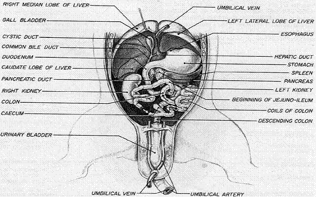
Thoracic Cavity: After leaving the stomach via the pyloric sphincter, food enters a short segment of the small intestine called the duodenum, which receives secretions from two accessory glands which contribute to the digestion and absorption of food, as well as secretions from the wall of the duodenum itself.
The first of these is the pancreas which is located in the mesentery running between the stomach and duodenum. It produces several digestive enzymes and buffers which empty into the duodenum via the pancreatic duct. The second is the liver, a large, multilobed, dark red organ filling much of the right anterior portion of the abdominal cavity.
It produces a yellow/green fluid called bile which is carried to the duodenum by the common bile duct. Although most mammals possess a bulb-like gall bladder which stores and concentrates bile, this structure is absent in the rat. Bile contains bile salts which facilitate the digestion and absorption of fats. Other prominent abdominal organs that you may wish to locate at this time are the spleen, kidneys, urinary bladder, and uterus. They will be described further in the dissection of the appropriate organ systems.
III. Questions/Data Collection
1. Be able to find the classic endocrine glands in the fetal pig and rat.
IV. Materials:
1. Dissection instruments and trays 2. Large size preserved fetal pigs, one per group 3. Several large live male and female rats 4. Ketamine/Xylazine/syringes 5. Heat lamps for rat surgery 6. Rubber gloves
V. References:
1. http://www.aug.edu/biology/ratlab1p.pdf
2. Walker, W.F. 1974. Dissection of the fetal pig, WH Freeman and Company
|