![]()
|
|
|
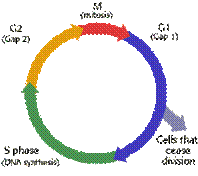
LABORATORY FIVE THE CELL CYCLE AND CELL DIVISION OVERVIEW: This lab involves a great deal of microscopy in order to demonstrate the processes of cellular reproduction, the basis for the reproduction of whole organisms. Dividing cells undergo an invariant sequence of events, which, though occurring in continuous succession, are recognizable as discrete stages. Today you will learn to identify the signs of these processes, and thus be able to recognize by name each of the phases of mitosis and meiosis. You will also draw these phases and, by counting their relative frequency, represent in a graph their approximate contribution to the complete cell cycle. Before looking at prepared slides of dividing cells, you will begin a simple exercise to stain chromosomes from the tips of onion roots. This procedure is not difficult, although it is imperative that you follow directions carefully. The staining procedure for staining the chromosomes takes time so that while you are waiting you can study the prepared slides for estimating the time that these cells spend in each phase of mitosis. Also be sure to see the large polytene chromosomes from larval insect salivary glands on prepared slides. OBJECTIVES OF LABORATORY: • To learn the stages of cell reproduction, their major events, and their sequence. • To understand the separation of chromosomes during mitosis and meiosis. • To be able to recognize mitotic phases in both plant and animal cells. • To collect and analyze data on the relative duration of phases of mitosis. • To study polytene chromosomes from insect larvae. • To stain chromosomes of onion root tips. INTRODUCTION: The division of nuclei by mitosis is exhibited by the somatic cells, all cells except the gamete producing cells, in most plants and animals. During its life span, a cell passes through a regular sequence of physiological events called the cell cycle. This sequence includes several distinct stages, each characterized by certain metabolic activities of the cell. In actively dividing cells, the cycle may last only a few hours; in other cells, the cycle may last for days or weeks. At the completion of the cell cycle a new generation of cells is produced. The cell cycle is made up of four phases: G1, S, G2, and M. Together G1, S, and G2 constitute interphase. Mitosis (M phase) follows the G2 phase and involves the division of chromosomes and the formation of two new nuclei. Mitosis consists of four distinct stages but actually takes up only about 5-10 percent of the complete cell cycle in most cells.
Cell Growth, Division, and Specialization. 26 June 2003 www.connect.ab.ca/~lburns/ cell_cycle.gif The duplication of the genetic material (DNA synthesis) in the nucleus occurs in the nondividing cell prior to the initiation of mitosis. The chromosomes are already doubled when they become visible during prophase, the first stage of mitosis. The term mitosis refers specifically to the process of nuclear division, the orderly distribution of the chromosomes between two daughter nuclei, starting with prophase and continuing through metaphase, anaphase, and telophase. Mitosis therefore occurs after duplication has been completed. Nuclear division usually occurs in close association with division of the cytoplasm (cytokinesis). The fact that these two processes do not always occur together indicates that different chemical and physical processes are involved in nuclear division (mitosis) and in cytoplasmic division (cytokinesis). An important point to remember is that cell division is a dynamic series of events during which the cell undergoes dramatic and often rapid physiological and morphological changes. The so-called stages of mitosis merely represent a few morphologically identifiable points in this continuum. Mitotic cell divisions are important in growing and regenerating regions of organisms. In plants, they are confined mainly to the meristems, such as buds, root tips, and cambium. In mature animals, mitotic divisions are found in cells at the base of epithelia, in the bone marrow, and in many other regions where tissues are growing or cells are being replaced. In animals, one place where dividing cells are easily located is in embryos. Meiosis, in contrast, is a mechanism for reducing the number of chromosomes by half. In animals, this takes place in cells that will give rise to gametes, but it may occur at other points in a sexual reproductive cycle. In plants, meiosis results in the formation of spores. To understand meiosis, recall that chromosomes commonly occur in pairs; members of a pair carry genetic factors for the same traits and are called homologous chromosomes. During mitosis, homologous chromosomes behave independently, and a copy of each is transmitted to each daughter cell. During meiosis, however, the homologues associate or synapse; synapsis is followed by separation of the members of each homologous pair into different daughter cells. This fundamental difference in chromosomal behavior—independence of homologous chromosomes in mitosis and association and separation of homologous chromosomes in meiosis—is the key to understanding the differences between these two otherwise mechanically similar processes.
STAINING CHROMOSOMES OF ONION ROOT TIPS. Start this part of the lab early in the lab period and then go to work on another section. The meristems, growing tips—shoot and root of plants, have numerous cells in division. In a few steps, you can make a slide that will show stages of mitosis in the root tips that have been growing in water for about a week. Using the following procedure, you and your partner should prepare a squash preparation of root cells that will show some cells in stages of mitosis. Find several onions at the front desk from which to take a root tip.
1. Cut a root segment at the base of the onion. Now, cut off the root tip about 1.5 cm long. Be sure that you are using a root tip—it should have a point on one end. If you don’t use a tip, you are not likely to find dividing cells. Put the root tip in a watch glass of Ethanol (EtOH)/acetic acid fixative for about ten minutes. It will tend to float on the surface so use forceps to push it into the solution.
2. Place the root tip in Ethanol (EtOH)/Hydrochloric Acid (HCl) for five to ten minutes. Put a top on the watch glass with this solution so that you will not breathe the fumes, and do not get the solution on you or your clothes. This solution dissolves the material between the cells. When the cells are free from one another, they will be more easily spread out into a single layer and be more easily flattened to reveal the chromosomes in the nucleus.
3. Place the root tip in the watch glass of Ethanol (EtOH)/acetic acid fixative for about a half an hour. Put a top on this solution.
4. Now, here is the delicate part. Put the root tip on a glass slide. Look at your root tip. There should be a pointed end, the tip, and a blunt end where you made the cut from the bulb. Using a razor blade, cut 1 mm (just one!) from the tip end—just the tip. Discard the rest of the root. On the tip, place a drop or two of acetocarmine stain. Let this stain sit on the root tip for ten minutes. Add more stain to keep the root tip wet. After staining, put a cover glass on the root tip and press gently using the bibulous paper book.
5. Heat the slide gently with an alcohol flame. The alcohol burner should be at your desk. Press out again using the bibulous paper book.
6. Examine the slide for chromosomes.
7. If you do not see dividing cells, you may need to add more stain. Do this by adding a drop at the edge of the cover glass. Wait, press out, and examine again.
8. Have an instructor check your slide.
If you are successful, you will see similar stages of mitosis as you see on the prepared slide of Allium, although this time you are looking at squashed out cells instead of sectioned material. If you are very successful, this would be the best way to count chromosomes. Why would that be true?
MITOTIC PHASES: This part of the lab involves the study of a slide of dividing cells in the root-tip meristem and the embryonic cells of the whitefish blastula. A clear understanding of chromosomal behavior in both mitosis and meiosis is essential to your later work in genetics. Obtain a slide of representative plant and animal mitotic tissue (onion root tip and whitefish blastula).
1. Observe your root tip preparation under low power and sketch the general features in the space provided below.
2. Once the cells are in focus, switch to high power and identify the different phases of mitosis and make drawings of each phase.
|