![]()
|
|
|
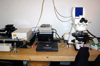
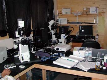
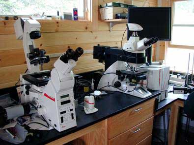
I have always been interested in imaging of biological specimens. I started doing light and electron microscopy in graduate school and in my post-doctorial training. In recent years I have shifted my attention to fluorescent microscopy and image processing. This is a very dynamic field and it is difficult to learn many of the techniques on your own. I have been lucky to have taken a couple of excellent workshops to help get me up to speed. The first was the Analytical and Quantitative Light Microscopy (AQLM) course offered by the Marine Biological Laboratory at Woods Hole (http://www.mbl.edu/education/courses/special_topics/aqlm.html). There is another workshop that I had been trying to get into for several years. It is very popular course and difficult to get into. After several years of applying I got in and was able to take an excellent workshop on Quantitative Fluorescent Microscopy at the Mount Desert Island Biological Laboratory in Maine (http://www.mdibl.org/courses/Quantitative_Fluorescent_Microscopy/159/). All the major microscope manufactures were there with their best fluorescent and confocal microscopes. Below are some of the imaging workstations I used and some of the images I collected during the workshop.
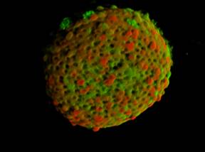
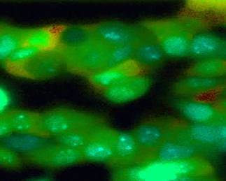
Zeiss Pascal Workstation 3D-Reconstruction of Islets cells In my opinion, the Zeiss optics and software were the best I saw at the workshop. The image above is a two-dye fluorescent staining of a group of cells of the Islets of Langerhans. The image above is 3-D confocal reconstruction of a series of planes of focus (Z-stack).
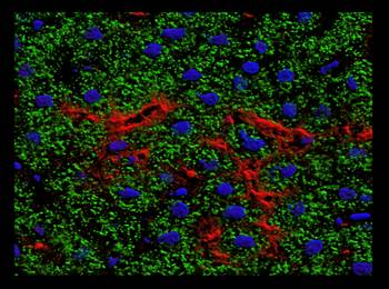
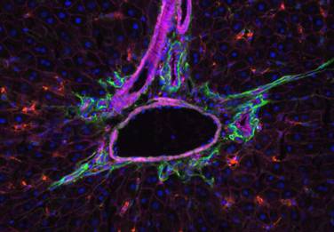
3-D Liver Reconstruction Blood Vessel in Liver Above are a couple more images I got from the Zeiss workstations. The one of the left is a 3-D reconstruction of a stack of confocal images using three dyes. The blood vessel on the right is a widefield fluorescent (not a confocal image) image of a blood vessel in the liver with three fluorescent stains.
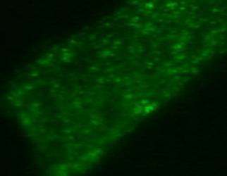
Olympus Workstations BTC-3 Cells loaded with Fura-2 Olympus had a series of microscopes, both widefield and confocal. All of them were motorized and most of the control of the scopes was computerized. One of the procedures allows for the visualization of intercellular calcium levels (Calcium-Ratio Imaging). The BTC-3 cells above loaded with the dye Fura-2 change color when cellular calcium is released.
TIRF Image of Membrane We also did a procedure called Total Internal Reflection Fluorescence (TIFR) microscopy. This is a procedure that uses the laser of a confocal microscope to image just at the plane of the cell membrane where the cell in in contact with the coverslip. It is possible to observe membrane activity not possible with other methods. I have a cool time-lapse movie of the TIFR activity I will try to get linked to the image above.
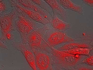
Nikon Workstation DIC/Fluorescent Overlay of CV-1 Cells Nikon also had multiple widefield and confocal microscopes available. I especially enjoyed working on the microscope workstation above because it used the same image-processing software I use, that is Universal Imaging's MetaMorph package. Above is one field of a timelapse experiment with CV-1 cells. The cells are imaged with both DIC transmitted-light and fluorescently with mito-tracker red dye. The transmitted light and fluorescent images are overlayed using the MetaMorph software.
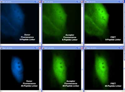
Leica Workstations FRET Images of CFP/YFP in Cos-7 Cells We did a number of activities at the Leica workstations. Above are some images from an experiment we did using a procedure that was new to me called FRET. FRET stands for Forster Resonance Energy Transfer. This is a procedure that allows you to visualize when two molecules have come within close proximity of each other in a cell. Different proteins are labeled with two different dye molecules. By themselves, the dye molecules do not fluoresce, but when they get close together, they produce a fluorescence you can see, way cool stuff.
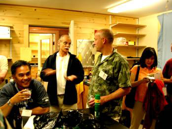
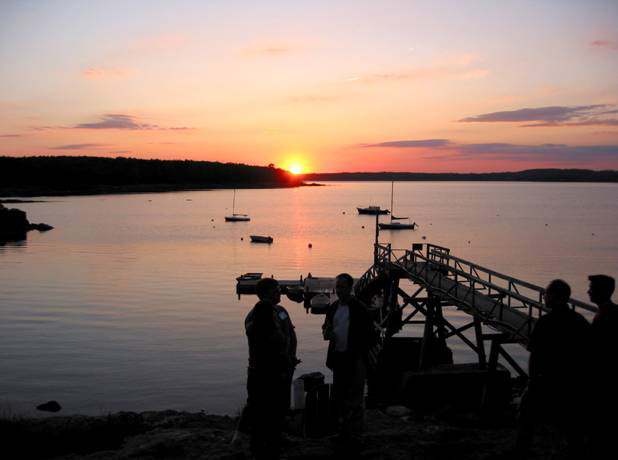
Simon Watkins Dave Piston Above are two of the faculty that taught the week-long workshop. Simon Watkins from the University of Pittsburgh was organizing the program and David Piston from Vanderbilt Medical school was coordinating many of the lab activities. If you have a chance, you should visit of attend workshops at the Mount Desert Island Biological Laboratory, great facilities, and not a bad location, at least in the summer months....
View out the back of the lab.... |