![]()
|
|
|
LAB 11: THE FETAL PIG Introduction In today’s lab we will examine the structure and function of a representative vertebrate, the fetal pig. We will be working in groups of two students and concentrate primarily on the circulatory system, the respiratory, digestive, and urogenital systems. It will be helpful to pay particular attention to the unified manner in which these organ systems interact to coordinate the animal's response to its environment. Your instructor will inform you of the terms and concepts for which you will be held responsible, although any words in boldface type are a good indication (spelling included).
Pre-Lab Questions Note, this is a lot of material, but it summarizes what you will be exploring in today’s lab.
1. Where are the Dorsal, Ventral, Medial, Lateral, Anterior and Posterior aspects?
2. Describe the regions of the digestive system that food must pass between the mouth and anus.
3. Describe the path an egg follows as it leaves the ovary.
4. Describe the path a sperm cell takes as it leaves the testis.
5. Describe the path between the aveoli in the lungs and the opening of the nostrils.
6. Describe the path blood takes as it passes through the majors vessels and chambers of the heart.
Procedures
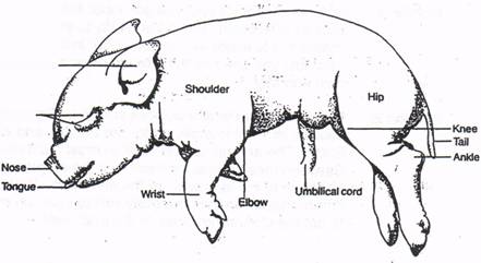
Thoroughly rinse your pig under cold running water right away to reduce the irritating preservative odor (don't worry—you won't wash out the preservative). As soon as you have made your initial incisions and opened the thoracic and abdominal cavities, rerinse your pig again. Examine the external anatomy of the fetal pig, including the openings of the reproductive, digestive and reproductive tract. What sex is your pig?
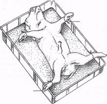
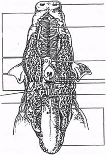
Once you are familiar with the external anatomy, tie it down to a dissecting tray as indicated in the figure on the left below.
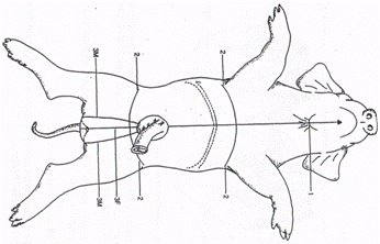
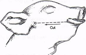
Insert the blade of your scalpel under the pig's chin, just below the surface of the skin, and cut ventrally to the umbilical cord. Be careful not to make the cut too deep; instead, make several shallow cuts until you can see the internal organs. Cut around the umbilical cord and stop.
Next, make two lateral cuts, as shown in the figure above. Begin at the midline, and cut laterally under the armpits and above the hind-limbs. The anterior lateral incision must cut through the rib cage.
Between these lateral incisions lies the diaphragm, which is attached to the side walls of the abdomen. You can cut the diaphragm away from the body wall to open up the cavity. Be sure to cut the diaphragm close to the body wall.
If you have a male pig, make two incisions (incision 3M) 2 cm on either side of the midline caudally to the tail. If you have a female pig, continue incision 1 to the genital papilla (incision 3F). When it comes time to do the dissection, dissect carefully and do not mutilate your pig. Use as much blunt dissection as possible (your instructor will demonstrate). You may often find scissors more useful than scalpels. Only remove organs when instructed to do so, or you may not be able to locate structures described later.
Do not wash any waste tissues down the drain - simply wrap them in paper towels and discard them in the garbage cans. Remember to play it safe in the lab: always handle dissecting instruments with extreme caution, and wear latex gloves at all times.
Read each section thoroughly; understand it completely before you start cutting. Try to find landmarks. Look for something before asking for help! The instructor's job is to guide and assist you in dissection and identification, not to find everything for you! You may find it helpful to examine the models and any other dissection manuals or atlases on display.
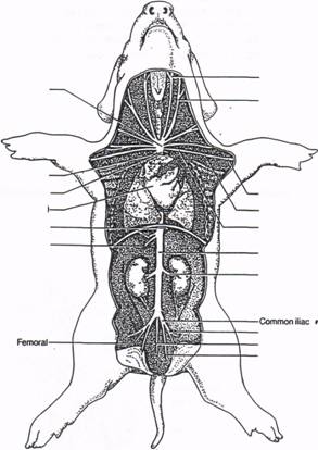
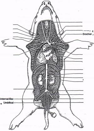
Remember throughout these dissections that left and right orientations refers to the pig's left and right, not yours. Not all pigs are identical. Aside from the obvious male/female dichotomy any differences in pigmentation and external anatomy, there may be anomalies ranging from the precise pattern of circulatory vessels to the exact arrangement of muscles. These pigs have been injected with latex so that arteries appear pink and veins blue.
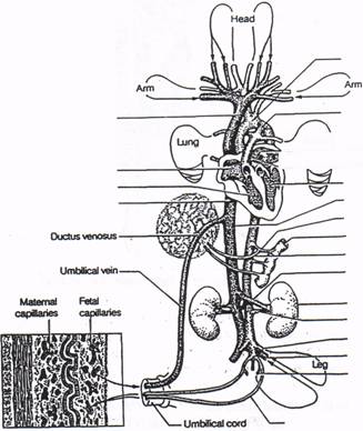
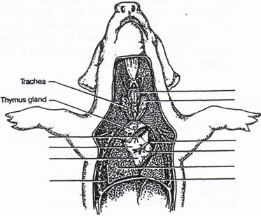
1. Once you have completed incisions 1 through 3 above, pull the skin flaps back to reveal the internal organs. Notice that the umbilical cord contains three vessels that lead into the abdomen: two pink umbilical arteries, which carry deoxygenated blood and wastes to the placenta; and a blue umbilical vein, which carries oxygenated blood to the heart. Between the umbilical arteries is the urinary bladder.
2. Before you cut the umbilical vein, tie a small length of string around the vein close to the liver (the dark brown organ occupying most of the abdomen and another length of string close to the site where the vein enters the umbilical cord. This procedure will help you find the vein later. As the umbilical vein enters the liver, it becomes the ductus venosus.
3. Now cut the vein between the two knots and pull the umbilical cord caudally. At this point, you may find it necessary to rinse the inside of the pig with running water. Your ultimate goal in this exercise is to locate the major vessels of the circulatory system. You will first examine the heart and its major vessels; then you will move on to the upper and lower veins; and finally you will study the upper and lower arteries.
Heart and Circulatory System
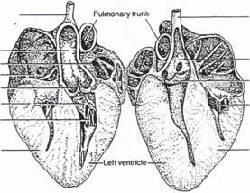
The dark flapiike structures on top of the heart are the right and left auricles, which are extensions of the atria. The greatest portion of the heart consists of the ventricles. Traversing the left ventricle is the left coronary artery.
Protruding from the right side of the heart and arching dorsally is the pulmonary trunk; this structure branches into the right and left pulmonary arteries, which lead to the lungs. Since the lungs do not function in the fetus, most of the blood is shunted away from the pulmonary trunk through the ductus arteriosus to the aorta.
Leading into the heart's right atrium are two large veins, the cranial and caudal vena cavae, which collect blood from the tissues of the upper portion and the lower portion, respectively, of the body. Below are figures of the heart, and major vessels of the fetal pig.
Respiratory System
1. Place your fetal pig dorsal-side-down on the dissecting tray, and secure it with string.
2. Use your scalpel to make two incisions, one on either side of the mouth, then pull the lower jaw down as you cut until the oral cavity is exposed.
3. Identify the external nostrils, which open into the nasal cavities. The roof of the mouth and the floor of the nasal cavity are separated by a ridged hard palate and a slightly more caudal soft palate.
4. Pull down on the lower jaw until you see the caudal end of the soft palate, with a large round opening into the back of the nasal cavity.
This opening is the nasopharynx. When you pulled your pig's lower jaw back, you probably saw a flap of tissue attached to the base of the tongue. This is the epiglottis, which covers the opening of the air passageway (trachea) when food or liquid passes over it to enter the esophagus.
5. Pull back on the epiglottis with your probe to see the glottis, which opens into the larynx.
6. a. Now close the pig's mouth, and probe the top of the neck to locate the larynx. You may have to cut the muscles surrounding the larynx to reveal this cartilaginous structure. Caudal to the larynx is the trachea (Figure 21.12).
b. Note the cartilaginous rings that support this tube.
c. Carefully follow the trachea caudally as it extends dorsal to the major arteries and the heart in the thoracic cavity.
7. With the approval of your instructor, you may remove the heart to reveal the trachea as it splits into right and left bronchi. The bronchi are also supported by cartilage.
The lungs are contained in the pleural cavity. This cavity is lined by a thin outer membrane, called the parietal pleura, that lies against the rib cage, and by another membrane (the visceral pleura) that encases the lungs. Together, these membranes form a sac filled with fluid that helps reduce friction as the lungs expand and contract.
The lungs and heart are protected by the rib cage. Between each rib are the intercostal muscles which expand and contract, alternately enlarging and compressing the rib cage during breathing. The lower border of the thoracic cavity is the muscular diaphragm you encountered before.
8. a. Examine each lung.
b. Count the number of lobes and record this number in item #3 at the end of this section.
9. Trace the left bronchus as it enters the lung and then divides into secondary bronchi. You will find it difficult to follow the respiratory tubes beyond the secondary bronchi. The lungs consist mainly of air sacs (currently fluid-filled), called alveoli, and as a result have a spongy texture.
10. Once you have completely examined the respiratory system of the fetal pig, answer the questions in item #3 at the end of this section.
Digestive System
1. Place the pig dorsal-side-down, and secure the limbs with string.
2. Examine the oral cavity as you did above, but this time focus on its role as part of the digestive system.
You can immediately see the tongue, which is lined at the front with papillae containing taste buds. You may also be able to see a few teeth protruding along the outside of the hard palate. The posterior end of the oral cavity is the pharynx, into which several tubes open. In addition to the dorsal nasopharynx, which leads to the nose, and the glottis, which opens to, the larynx, there are two Eustachian tubes (one leading to each ear) and an esophagus, which travels to the stomach.
3. a. Pull the lower jaw down far enough to expose the opening of the esoph- agus.
b. Move a blunt probe down the tube.
c. Distinguish the esophagus from the glottis.
Once food is chewed, the tongue pushes the food bolus to the back of the oral cavity where it passes over the epiglottis and down the esophagus. Wavelike contractions (peristalsis) of the esophagus move the food into the stomach.
4. Look for the esophagus from the direction of the thoracic cavity, where it lies dorsal to the trachea.
5. Trace the tube caudally as it penetrates the diaphragm and ends at the stomach. Cut open the stomach to see the rugae — ridges within the mucosa that allow the organ to expand.
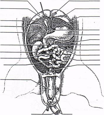
6. At the proximal end of the stomach is the pyloric sphincter, a valvelike muscle that controls the passage of partially digested food into the small intestine. The small intestine is a long, convoluted tube within which the digestive process is completed and nutrients are absorbed. The pancreas lies between the stomach and the first portion of the small intestine, known as the duodenum. The large organ in the upper right quadrant of the abdomen is the liver.
7. Lift the liver and you will see the saclike gall bladder, which is drained by a tube called the cystic duct. Bile stored in the gall bladder flows through the cystic duct and enters the duodenum, where it aids in the digestion of fats. Dorsal to the stomach is a dark, finger-shaped organ, the spleen.
8. Follow the small intestine until it joins the large intestine. The union of the two tubes is the site of another sphincter, the ilegcecal yalve. Adjacent to this valve is a short, blind pouch, the caecum. The large intestine, also called the colon, is much shorter than the small intestine and is less coiled.
9. Follow the colon as it passes along the dorsal wall of the abdomen; this portion of the tube is called the rectum. At the terminal end of the rectum is another sphincter muscle, the anus, which is located at the base of the tail.
The abdomen itself is known as the peritoneal cavity. The thin outer lining of the cavity is called the parietal peritoneum, while the membrane lying directly on the abdominal organs is called the visceral peritoneum. As occurs in the pleural and pericardial cavities, these membranes form a sac that is filled with fluid.
Excretory System
The kidneys lie dorsal to the small intestine. You will see a thin overlying tissue — this is the parietal peritoneum.
1. Cut this tissue away. The curvature of each kidney faces the midline, and the rectum and the dorsal aorta pass between them. Three tubes emerge from the kidney: a renal artery; a renal vein; and the ureter, which collects urine and carries it to the urinary bladder.
2. Trace the ureter until it reaches the bladder. If you have a male pig, the animal's urethra proceeds ventrally and then curves toward the umbilical cord. The thick tissue around the distal end of the urethra is the fetal penis. 3. Cut the ureter close to one kidney and remove the organ.
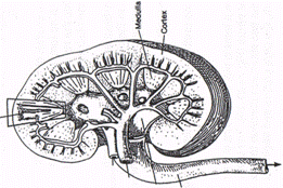
4. With your scalpel, slice the kidney lengthwise so that it looks like the drawing above.
This sagittal section of the kidney reveals the outer renal cortex and the inner medulla. Urine formed in the nephrons is collected in the collecting ducts and funneled into the renal pelvis. From the pelvis, urine flows into the ureter and thence to the bladder.
The urinary bladder lies between the umbilical arteries. Its medial end is connected to the ureters, which arrive from the kidneys, and to the urethra. The urethra drains urine from the bladder to the outside.
5. Locate the urethra.
6. To follow the urethra, cut through the pelvic bone and spread the hind limbs apart. If you have a female pig, the urethra is short and joins the vagina to form the urogenital sinus. The urethra lies ventral to the vagina, and in a living pig the two structures continue to separate as the fetus matures. By the time of birth, the pig's urethra and vagina are completely separate t ubes.
7. When you have completed this exercise, answer the questions in item #5 at the end of this section.
Reproductive System
The term urogenital implies that the reproductive structures are closely associated with the excretory structures. You will now return to this area to examine the reproductive structures in more detail.
Female reproductive organs: Just posterior to the kidneys lie the female gonads, the ovaries. Each ovary is kidney-shaped and about 5 mm long. A minute oviduct attaches to the ovary; it is the tube through which eggs pass to the uterus. The uterus in turn has two long, hornlike processes, one passing to each ovary. The central body of the uterus lies medially and just dorsal to the urethra.
1. Follow the uterus caudally to where it joins with the vagina and eventually unites with the urethra to form the urogenital canal. Within the ventral surface of the canal lies the clitoris, a small protuberance homologous to the penis in the mate.
Male reproductive organs: The location of the male gonads, the testes, in a mammal depends on the animal's age. In young pigs, the testes are in the lower abdomen posterior to the kidneys. As the pig matures, the testes drop into the scrotal sacs, which are ventral to the anus. As a testis descends into the scrota) pouch, a passageway (the inguinal canal) forms. This canal contains blood vessels, nerves, the spermatic cord, and the sperm duct.
2. With your scalpel, cut open a scrotal sac to reveal the testis, which is usually dark brown.
Adjacent to the testis is a lighter-colored tube, the epididymis that begins at the cranial end of the testis and ends caudally. The epididymis then continues as a smaller tube, the vas deferens that carries sperm from the gonads internally through the inguinal canal and passes to the medial end of the urinary bladder.
3. Cut the ureter on one side of the bladder and pull the bladder ventrally.
You will see that the two sperm ducts join to form an ejaculatory duct. As the ejaculatory duct descends, two small white glands, the seminal vesicles, appear. These glands provide a nourishing seminal fluid to surround, maintain, and transport the sperm.
The ejaculatory duct joins the urethra and, during copulation, transfers sperm from the penis to the vagina of the female. On both sides of the urethra, just dorsal to the pubic bone, are two long, slender Cowper's glands. These glands add seminal fluid to the ejaculate. The urethra continues ventrally through the penis where it opens just posterior to the umbilical cord.
Materials
Dissecting Trays Probes, dull probes, scalpel, scissors Razor blades Dissecting pins Fetal pigs, one for each 2 students String to tie pigs to trays
|