![]()
|
|
|
LAB 8: ANIMAL DEVELOPMENT
Introduction Animal development is a complex process of sequential activation of genes, which in turn code for proteins that allow for the growth and differentiation of the organism. Like many topics we examine in the lab, this is designed to be an introduction to selected aspects of the field of development. We will examine prepared slides as well as two live animal models to get a sense of the changes that occur during early development. Pre-Lab Questions 1. What Phyla and Class are sea urchins, Zebrafish and chicks found?
2. What is a Blastula and what is a Gastrula?
3. What is Ringers Solution?
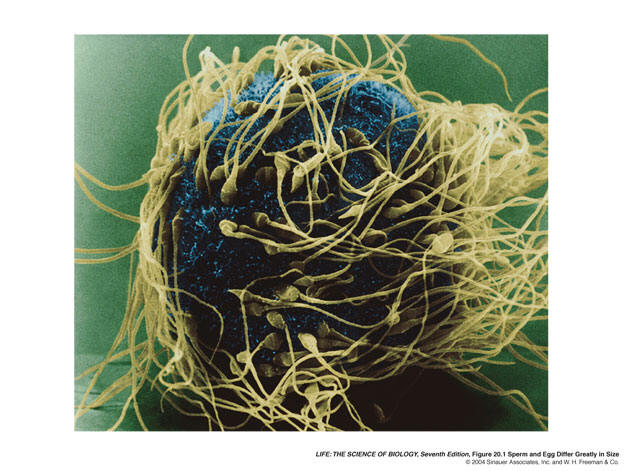
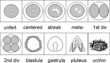
Procedure A. Sea Urchin Development Cleavage in the sea urchin proceeds in a predictable fashion. The first two cleavages are meridional (longitudinal) and the third is equatorial, giving rise to eight equal-sized cells or blastomeres. The sea urchin eggs have an animal pole and a vegetal pole that define the animal/vegetal axis.
Obtain a slide of Echinoderm development and make drawings and identify the: 1. Early cleavage stages 2. Blastula,and Blastocoele 3. Gastrula-Stage Embryos 4. The Primitive Gut or Archenteron
Early Cleavages Blastula Stage Gastrula Stage
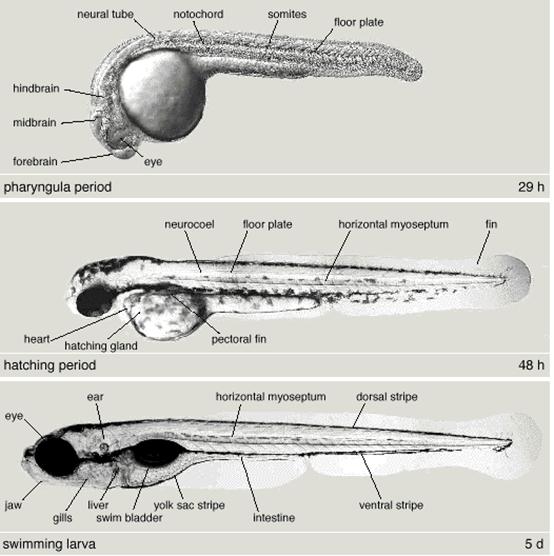
B. Zebrafish Development With luck, our breeding colony of zebrafish will provide embryos that will be about 6 hours old. The zebrafish is a wonderful model organism in which to study development. A classic paper on zebrafish development was done by Kimmel, et al in 1955 (Kimmel et al., 1955. Developmental Dynamics 203:253-310.). We will use his original sketches of zebrafish development to help us understand what we are seeing in the embryos.
The procedure involves getting a dissecting microscope and placing several zebrafish embryos in a small (35 mm) Petri dish with at least 5mm of water. The embryos can then be observed over time through their transparent egg membrane or chorion. If you get your embryos soon enough, you should be able to identify the early cleavage and blasutla stages. If you have later stage embryos, say over 6 hours old, you should be able to see the gastrula stage. Once the generation of form or morphogenesis and the formation of the organs or organogenesis has occurred, you should be able to identify a number of structures including the: optic cup, brain regions, somites, otic vesicle, heart, tail bud and of course the yolk sac. Use this link to get more information and establish an age for each of the stages above, http://zfin.org/zf_info/zfbook/stages/stages.html
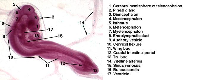
C. Chick Development Obtain slides of 72 hour old whole-mounts of chick embryos. These prepared slides will serve as a guide to find the structures in the live embryos which will be the same size. The structures are much easier to see in the prepared slides, because in vivo, or in the live embryo, the yolk sac obscures much of the detail of the embryo. Use manuals provided to help to identify major landmarks in the embryos including the: Head/Brain Regions Eye/Optic Cup Otic Vesicle Somites Limb Buds Heart Vitelline Vessels Before you obtain your fertilized egg, check all your equipment to be sure you have everything in readiness. Each group of students will be provided with a number of 72 and 96 hour old embyos. Once you are comfortable with finding structures in the prepared slides, you can obtain a fertilized egg. When getting an egg from the incubator, be sure to hold it in exactly the same orientation as it had in the tray, as the embryo is normally at the uppermost part of the egg and we want to be able to find the embryo. Pour about 1cm of warm sterile Howard's Ringer (HR) solution, a physiological saline solution, into a fingerbowl, then crack the egg on the side of the bowl. Carefully lower the egg into the Ringer's solution. The object is to not break the yolk. If the yolk breaks at this point, try to remove the vasculated area, that is the area with the blood vessels to a large petri dish filled with warm HR. If you did not break the yolk you should be able to see the embryo with its heart beating using the dissecting microscope. If no embryo is visible, use your finger to try to flip or roll the egg over to expose the embryo. It is essential that you do not allow the embryo and yolk to dry out. Apply several drops of warm sterile HR as necessary. The area of the yolk covered with blood vessels is called the area vasculosa. At this stage of development the blood in the vessels picks up oxygen and food for the embryo and disposes of embryonic waste. As the embryo grows this area increases. Refer to your atlas for a diagram of the chick. Use your light to keep the embryo warm, and continue to keep it moist with HR. Identify other prominent structures. Is the heart rate slowing down? If so, why?
Materials Live Chick embryos 72 and 96 hour old Zebrafish Slides of Sea Urchin development, chick embryo development Water bath set at 40oC for Howard's Ringers, or Ringers maintained in 40C incubator 6 liters of Sterile Howard's Ringers at 40C 37oC incubator with an open tray of water to maintain a high humidity for chick eggs sharpies pipettes with pumps Watchmakers forceps, regular forceps, and fine scissors 72 and 96 hour old live chick embryos filter paper and pasture pipettes and bulbs 1% solution of Neutral Red Inverted phase-contrast microscopes Squirt bottles for each group with 70% EtOH 24 inch white place mats latex gloves Pasture pipettes and bulbs Modeling Clay for egg nests Whole-mount slides of Chick Development Models of Echinoderm and Amphibian Development high intensity light sources for dissecting scopes large waste container for used eggs and Ringers Large watch glasses to hold chick eggs 35 mm Petri dishes for zebrafish embryos Howard's Ringers (2000 ml) NaCl 14.40 g CaCl 2H2O 0.46 g KCl 0.74 g Distilled H2O to 2000 ml Mix up and autoclave to sterilize ml Mix up and autoclave to sterilize
References 1. Jakoby, W. B. and Pastan, I. H. 1979. Cell Culture. Methods in Enzymology Volume LVIII. Academic Press, New York. |