![]()
|
|
|
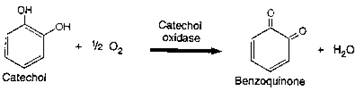
LAB 3: ENZYMES Introduction Virtually every chemical reaction in organisms involves enzymes. This lab will examine function of the enzyme catecholase (also known as tyrosinase). This is the basic enzyme-mediated reaction we will study in today’s lab:
Catechol is a derivative of benzene found in many fruits and other plant structures. Catecholase catalyzes the reaction of catechol and oxygen and is the enzyme that causes bruised or otherwise damaged fruit to turn brown. In the presence of catecholase, catechol is oxidized to form benzoquinone, which has a reddish brown color.
Pre-Lab Questions:
1. Define Enzyme
2. Define Substrate
3. Define Cofactor
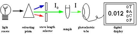
In order to observe the enzyme-mediated reaction we need an instrument that can measure the change in color from clear to brown. We have such an instrument called a spectrophotometer that works like this:
Procedure You will again be working in groups of two in today’s lab. All groups will first need to answer the following question: Question 1 – What is the rate of the conversion of catechol to benzoquinone by catecholase? You will be provided with the following materials: Spectrophotometer set to 360 nm 3 spectrophotometer tubes in rack with marking pencil 40º C water bath (or hotplate); 1 percent catechol stock solution catecholase stock solution in ice bath; distilled water As you need to be able to get good data on this reaction to be able to compare the action of the enzyme-mediated reaction under a variety of conditions, here are some steps to help you answer the question: Label test tubes 1, 2, and 3, and place 3ml of each of the following solutions into the respective tubes (each test tube will contain a total volume of 6 ml): Test tube 1: catecholase (always keep on ice!) and catechol Test tube 2: catecholase and distilled water Test tube 3: catechol and distilled water Cover the tubes with Parafilm and gently invert them to mix contents. Record the absorbency at 360 of each solution in item A of your worksheet. Place the three test tubes in a 40º C water bath for 15 minutes. Agitate the solutions and check the color intensity of each one when you begin and every 5 minutes thereafter. Read the absorbency in the spectrophotometer at 360. Record your data in the worksheet provided. At the end of the 15 minutes, remove the test tubes from the water bath. Save the tubes for color comparisons in later exercises.
Question 2 – How does enzyme concentration affect enzyme activity? You can vary the enzyme’s concentration by diluting it with a pH 7 buffer solution. A buffer solution resists any change in pH by releasing or taking up hydrogen ions (H+). Before you begin your experiment have your protocol approved by your instructor. You will be provided with the materials listed above for the basic enzyme reaction in addition to answer this question you will have: Additional spectrophotometer tubes 10 ml graduated cylinder; catecholase stock solution pH 7 buffer solution Notes: Record your hypothesis and procedures (approved by your instructor!) here:
Once you have made your solutions and noted the initial color intensities, place the tubes in the water bath for 15 minutes. Always keep catecholase on ice! Agitate the tubes every 5 minutes, and record the resulting color intensities. Record your results and conclusions, you can use Table B below to help.
CAUTION: Catechol, hydroxyquinone, and phenylthiourea (PTU) are hazardous if ingested or absorbed through the skin. If you are using these materials please wear plastic gloves when you handle the chemicals. If an accident occurs, immediately wash your hands thoroughly with warm water and soap. Question 3 – How does substrate concentration affect enzyme activity? That is, if you keep the amount of enzyme constant, how will the enzymatic activity change as you add more and more substrate? In addition to the materials available for the basic reaction above you will be provided with: Additional spectrophotometer tubes 10 ml graduated cylinder; 1 % catechol stock solution distilled water pH 7 buffer solution Notes: Record your hypothesis and procedures (approved by your instructor ) below:
Once you have prepared the solutions and recorded their initial color intensities, place the test tubes in the water bath for 15 minutes.
Agitate the tubes every 5 minutes, and note their color intensities.
Record your results and conclusions, you can use Table C below:
Question 4 - How specific are enzymes in terms of substrates? The specificity of an enzyme is that enzyme’s tendency to react only with a particular substrate. In this experiment, you will demonstrate that catecholase can distinguish between two quite similar compounds, catechol and hydroxyquinone. In addition to the materials available for the basic reaction above you will be provided with: 1 % hydroxyquinone stock solution Notes: Label test tubes 1 and 2. Place 3 ml of catechol in test tube 1. Place 3 ml of hydroxyquinone in test tube 2. Pour 3 ml of catecholase into each tube. Place the caps on the tubes, and shake the tubes to mix the solutions. Note the color intensities, and place the tubes in the water bath. Agitate the tubes every 5 minutes for 15 minutes, and once again note the resulting color Intensities by taking absorbency readings on the spectrophotometer. Record your results and conclusions, you can use Table D:
Question 5 - How does temperature affect the activity of enzymes? Enzymes, like all proteins, have specific three-dimensional shapes. The molecules are held in these shapes—which are critical to the enzyme’s proper functioning—by hydrogen bonds and other fairly weak bonds. Extreme heat can break the bonds and thereby change the enzyme’s shape. In addition to the materials available for the basic reaction above you will be provided with: 0oC, 40ºC, 60ºC and 80ºC water baths or hot plates Thermometer; 4 each 400-ml beakers; test tube clamps Notes: Record your hypothesis and methods (approved by your instructor) below:
Once started, your tests should run approximately 15 minutes each. Agitate the solutions every 5 minutes and note the resulting color intensities.
Record your data and conclusions, you can use table E below:
Question 6 - How does pH affect the activity of enzymes? Many of the chemical bonds that hold a protein in its three-dimensional shape are affected by the presence of hydrogen ions (H+). The buffer solutions in the series you will use in this exercise vary in pH. By exposing the enzyme-substrate complex to a range of pH values, you can test the effect of hydrogen ion concentration on enzymatic activity. In addition to the materials available for the basic reaction above you will be provided with: pH buffer series (including pH levels of 2, 4, 6, 7, 8, 10 and 12) Notes: List your experiment and procedural steps and have your hypothesis approved by your instructor.
As in the previous exercises, agitate the solutions at the beginning of the test and every 5 minutes thereafter for 15 minutes.
Note the resulting color intensities on the spectrophotometer.
Briefly state your results and conclusions, you can use Table F:
Question 7 - What role do cofactors play in enzymatic activity? Enzymes often (but not always) require “helper” substances to bind substrates and catalyze a reaction. These substances, called cofactors, generally occur in two forms: mineral elements and organic coenzymes. In this exercise you will explore a cofactor that catecholase needs to catalyze the reaction that turns catechol (and oxygen) to benzoquinone. In addition to the materials available for the basic reaction above you will be provided with: phenylthiourea (PTU) crystals; chemical spatula or lifter (Note: Since PTU is a hazardous chemical, your instructor will dispense it.)
Notes: Label two test tubes 1 and 2. Pour 3 ml of catecholase into each tube. To test tube 2, have your instructor add three or four PTU crystals. [Note: Too much PTU makes the solution opaque (cloudy), which can give a falsely high absorbance reading.] Screw on the test-tube caps or cover with Parafilm and shake tubes for 4-5 minutes. Add 3 ml of 1 percent catechol to each test tube. Shake the tubes briefly, and record the color intensities of the solutions. Place the tubes in the water bath for 15 minutes. Record the resulting color intensities as in the previous experiments. Record your data and conclusions and answer the following questions:
What happened in the test tubes in which phenylthiourea (PTU) crystals were added?
Can you explain your results? What relation does PTU have to catecholase?
Why do enzymes have cofactors? Are cofactors required for adaptive (“good”) evolutionary reasons, or merely for historical (“indifferent”) evolutionary reasons?
Materials Enzymes/Substrate: 300 mL catechol stock 1% **POISON** KEEP ON ICE 200 mL tyrosinase stock (kept in freezer) KEEP ON ICE 25 mL hydroquinone soln 1% (shelf) Phenylthiourea crystals Waste container Buffers at pH 2, 4, 6, 7, 8, 10, 12 (30mL each), More of pH 7 (150 mL) Thermometers 10 mL grad cylinders spec 20 spec 20 tubes test tube racks test tube clamps 400 mL beakers spatulas sharpies parafilm scissors water baths at 40 degrees C, and 60 degrees C hot plate at 80 degrees C plastic pipets 5 ml pipets with pipet pumps Ice
REFERENCES/SOURCES: Helms, D.R., C.W. Helms, R.J. Kosinski, and J.R. Cummings. 1998. Biology in the Laboratory, 3e. New York: W.H. Freeman.
| |||||||||||||||||||||||||||||||||||||||||||||||||||||||||||||||||||||||||||||||||||||||||||||||||||||||||||||||||||||||||||||||||||||||||||||||||||||||||||||||||||||||||||||||||||||||||||||||||||||||||||||||||||||||||||||||||||||||||||||||||||||||||||||||||||||||||