![]()
|
|
|
I have been interested in using the zebrafish as a model to investigate the role of retinoic acid on vertebrate development. Retinoic acid is a derivative of vitamin-A which has profound effects on the development of vertebrate embryos. These effects include the differentiation or formation of anterior structures of the nervous system including the eye. Much of retinoic acid’s action is mediated through a group of receptor proteins in the cell cytoplasm of the developing embryos. When retinoic acid binds to these receptors, the complex has the ability to bind to, and activate specific groups of genes. The protein products of those genes result in further development of the embryo. In order to do this work, I first needed to get a zebrafish colony going.
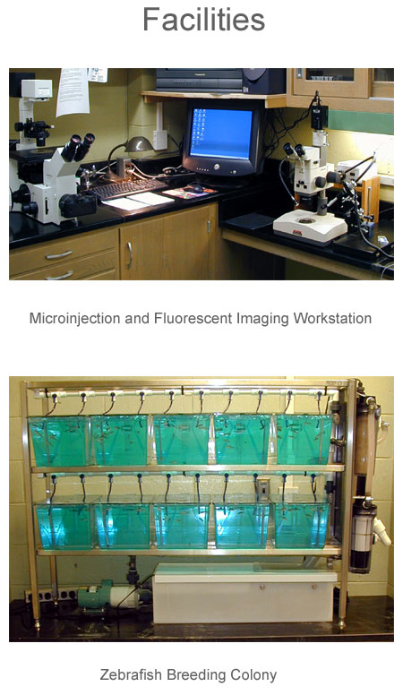
Zebrafish are one of the “sweethearts” of developmental biologists because of their small size and clear membranes (chorion) surrounding the embryo. It is necessary to get a productive colony of Zebrafish to provide a reliable supply of embryos. Stocks of breeding Zebrafish and a special protein-rich diet designed to maximize egg production were obtained. The conditions in the lab including were optimized for egg production. This included adjusting the water quality, temperature and photoperiod. Great care is taken with both obtaining and maintaining the breeding stocks, as the experiments can not proceed without embryos. Once the breeding system and conditions were optimized (seen figure of breeding system), large numbers of eggs were available on a fairly regular basis. For this study, I needed very early embryos, typically at the 1-2 cell stage of development. These early embryos were harvested first thing in the morning after the lights are turned on. In this phase of the project, one-celled Zebrafish embryos were obtained, staged and placed in special coverslip slide chambers for observation. MetaMorph image analysis software was utilized to control a Roper digital camera on the microscope. A series of still images were obtained of early developing embryos and the images were assembled into a time-lapse movie sequence. This procedure was necessary to eventually be able to track the migration fluorescently labeled cells. There were several challenges developing the protocols for photographing the embryos over a two day period, but the final images turned out very well. CD-ROMS of the time-lapse sequence of Zebrafish normal development generated are available upon request.
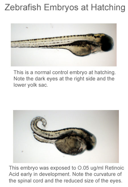
One of the long-term objectives of this study is to characterize cell migration in the presence of elevated retinoic acid concentrations. It was therefore first necessary to determine a range of retinoic acid concentrations that do not result in developmental abnormalities. In this part of the study, embryos were exposed to a graded series of retinoic acid concentrations for time periods of 12 to 96 hours. At elevated concentrations of retinoic a number of characteristic malformations were recorded.
These included curvature of the embryonic axis and edema, or fluid accumulation in the pericardial coelom, the sac that surrounds the heart. Based on these experiments two parameters were determined. The first is a measure of acute toxicity called the LC50 or lethal concentration to 50% of the population. The second was a working concentration of retinoic acid that did not result in any morphological anomalies. This working concentration of retinoic acid is being used in ongoing experiments.
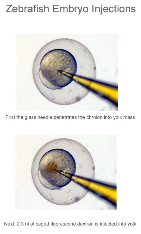
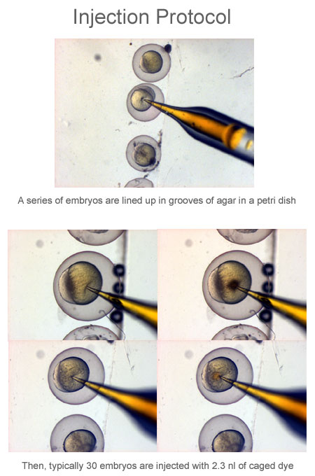
For years developmental biologists have labeled groups of cells in early embryos and then followed the developmental fate of those cells. In order to track groups of cells in the embryo some form of dye must be applied to the cells of interest. The dyes used to track cell movement in this study are fluorescent dyes that must be delivered to the embryo through the process of microinjection. In this part of the study, one to two-celled embryos were injected with different volumes and concentrations of normal fluorescine (not caged fluorescine) dye.
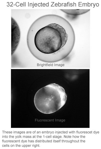
The dye was injected into the yolk sac of the embryo. In early embryos, many large molecules can move from the yolk sac into the cytoplasm. It was necessary to determine if injected dye could partition itself throughout the embryo. This stage of the study was successful and a procedure was developed that allowed all of the embryonic cells to be loaded with dye following injection into the yolk sac (See enclosed figures). The injected fluorescent dye was able to partition itself into all the embryonic cells of later-stage embryos.
If fluorescent dyes are to be used to track specific populations of cells in an embryo, the dye molecules must be introduced to a very small population of cells. It is very difficult to produce a needle small enough to inject a group of cells in a later (gastrula-stage) embryo. Such microinjection equipment is available, but it is very expensive and the procedures are quite complex.
An alternate procedure is to use “caged” fluorescent dyes. Caged fluorescent dyes are those that do not fluoresce unless they are activated by a laser or short wavelength UV light. In this procedure all of the cells of the embryo are loaded with caged fluorescent dye by microinjection into the yolk sac at the 1-2 cell stage. All subsequent cells in the developing embryo should contain the caged dye. The embryos were then allowed to develop to the late gastrula stage and a small group of cells were exposed to short wavelength UV light from the high magnification objective lens.
The 100X objective lens produces a very small point of illumination with high energy UV light which is sufficient to activate or uncage the fluorescent dye molecules in that specific group of cells.A series of experiments were performed and the process of loading the embryo with caged dye and uncaging specific groups of cells to track their migration worked well.
Unfortunately, the high-magnification lens of the inverted microscope produced a spot of UV illumination that larger than expected and it ended up uncaging too large of an area. Work is being continued to do not provide the resolution necessary to track small groups of cells. This protocol is still being developed. This work was been supported by HSC Summer Research Fellowships and VFIC Mednick Memorial Fellowship
|