![]()
|
|
|
I spent the Fall semester of 2007 in the lab of Dr. Nanette Nascone-Yoder in the Department of Molecular Biomedical Sciences, College of Veterinary Medicine, North Carolina State University. My work was supported by a ROA (Research Opportunity Award) supplement to Dr. Nascone-Yoder's NSF grant. Dr. Nascone-Yoder's lab has been interested in the development of the gut which starts off as a straight tube, then starts to from loops, and it the case of Xenopus, rotate. Most of the work in their lab has been with Xenopus, I was brought on board to use the zebrafish as a model organism to investigate gut development. One of the things that interested me about this project was the availability of zebrafish embryos as the NCSU lab has a good, dedicated animal-holding facility for experimental animals.
Front of the School of Veterinary Medicine at NCSU
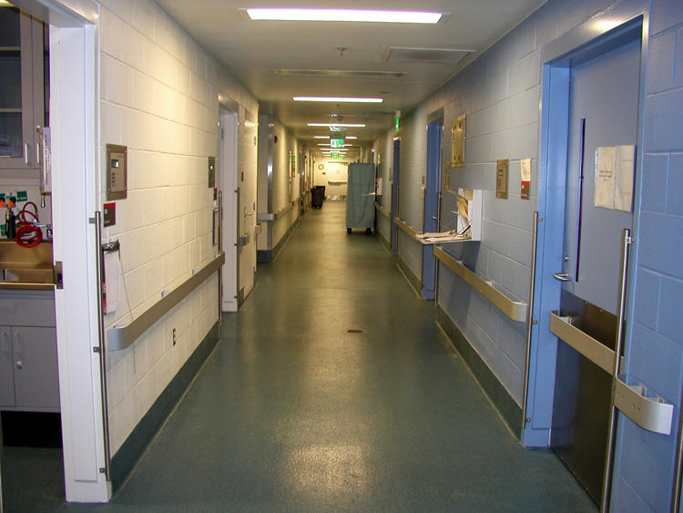
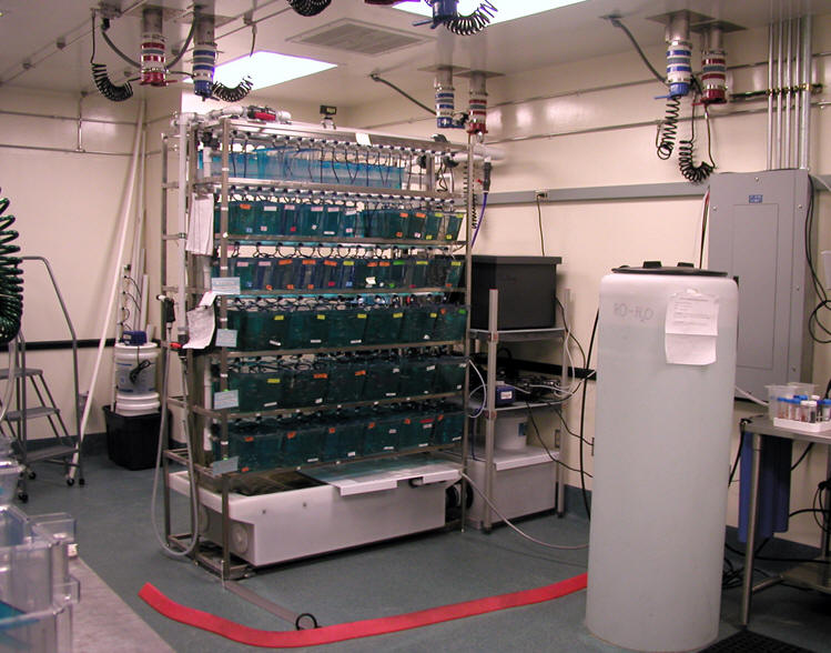
Hallway of animal-holding facility The Zebrafish Breeding Room
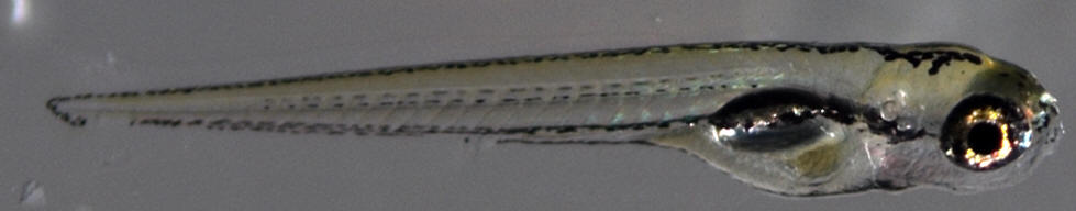
This is a view of a normal 5-day post-fertilization Zebrafish larvae
There are a number of projects going on in Dr. Nascone-Yoder's lab, but much of the work involves chemical genetic approaches to deconstruct the intracellular signals necessary for normal gut development. Chemical genetics is an approach that uses small, cell-permeable, biologically-active compounds which are know antagonists or agonists for specific molecules in a given signaling pathway. I was interested primarily activity of four molecules and how they influence gut development:
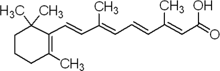
Retinoic acid is an endogenous molecule whose levels are spatially and temporally regulated during normal embryonic development via enzymes such as RALDH2 which synthesizes active forms or CYP26A1 which breaks down active forms.
Rockout is a cell-permeable ATP-competitive inhibitor of Rho kinase activity. It is a competitive inhibitor, yet it does not directly inhibit the activation of Rho Kinase.
Blebbistatin is also a cell-permeable compound that acts as a selective and reversible inhibitor of non-muscle myosin II. It has a number of activities including the disruption of cell migration and cytokinesis.
DEAB (diethylaminobenzaldehyde) is a cell-permeable compound that functions as an inhibitor of retinoic acid synthesis.
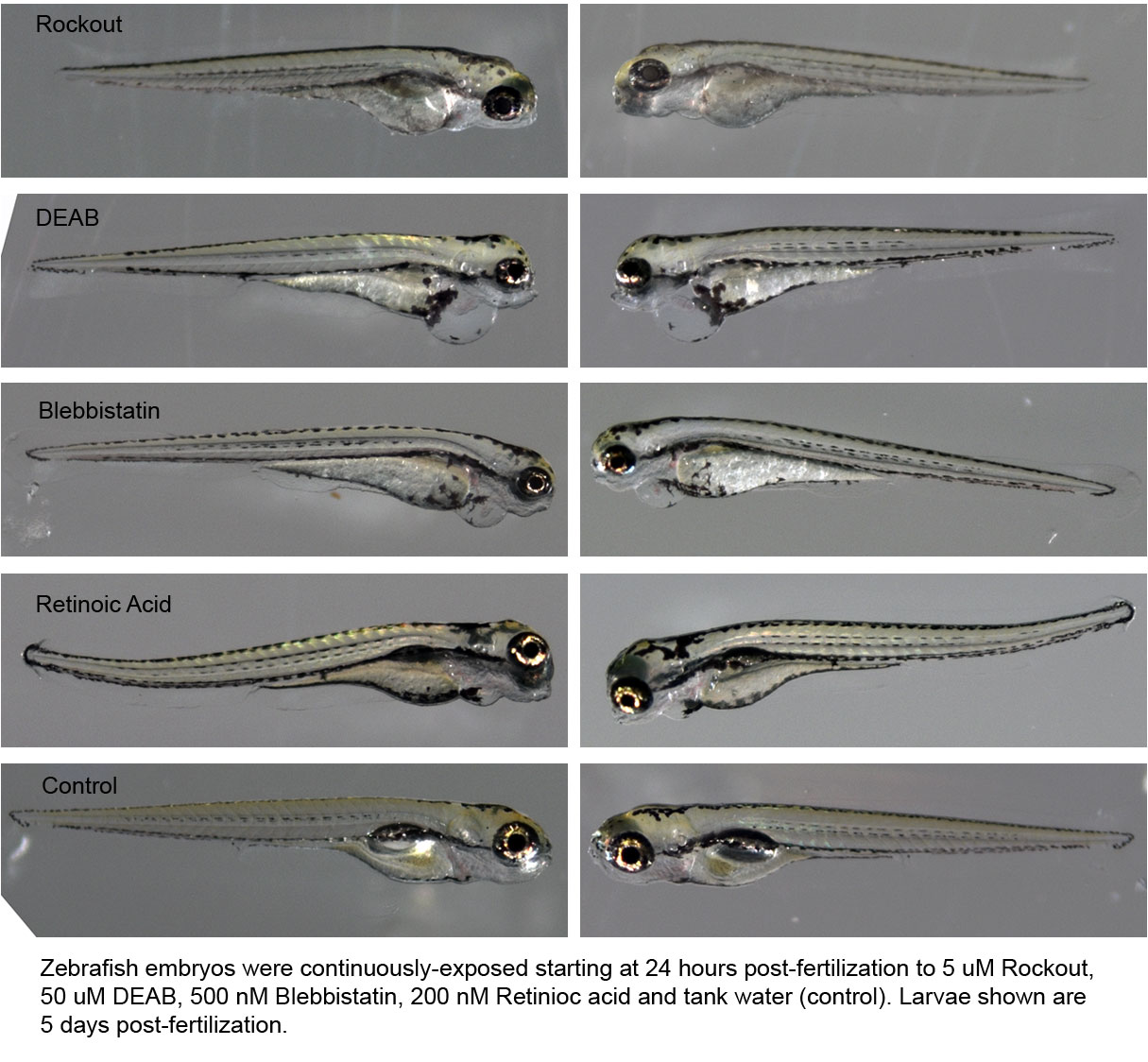
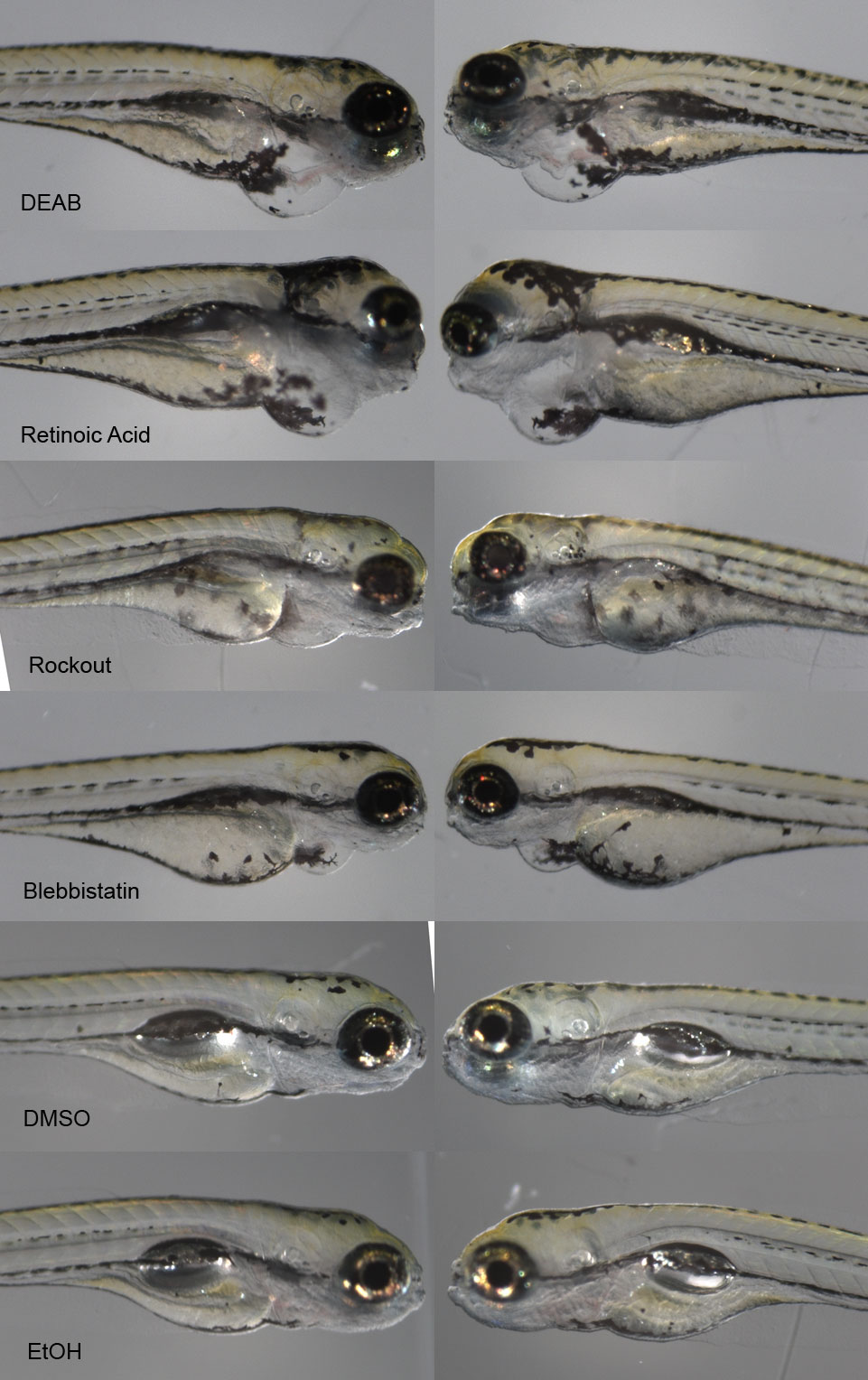
I needed to first determine a safe range of concentrations for the embryos and note their effect on the phenotype of the embryos and larvae. Below are several composite images of 5-day post-fertilization larvae that have been exposed to the compounds.
Below is another view
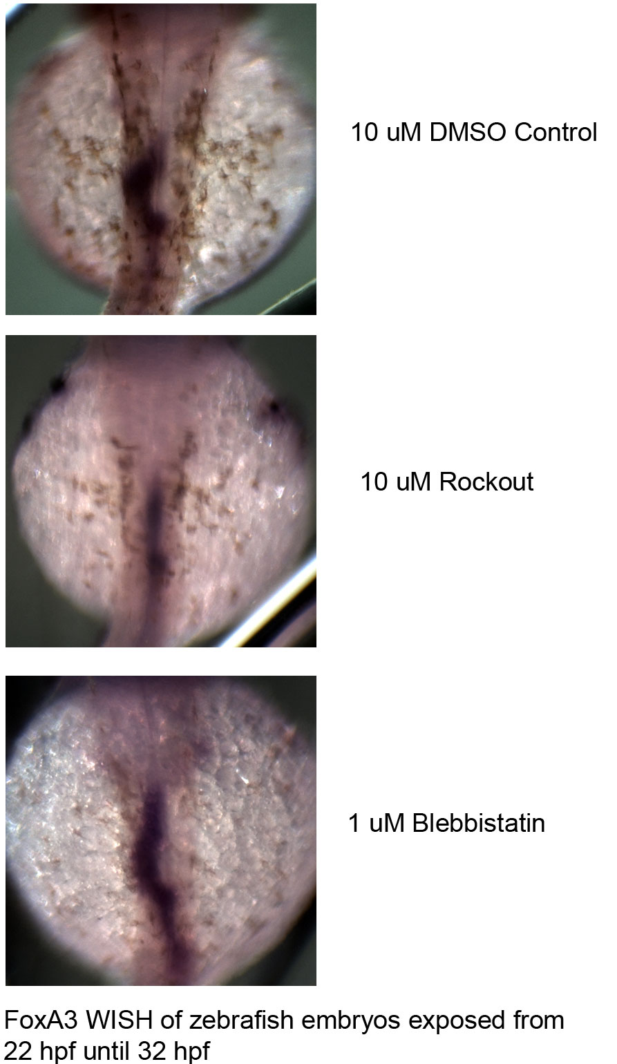
Once the range of exposure concentrations were determined we wanted to look more closely at what happens during early development when the gut first starts to loop in the zebrafish, about 24 to 30 hours post-fertilization. I used a procedure called whole-mount in situ hybridization or WISH. This procedure involves first making a DIG-labeled mRNA probe called Fox A3 (provided by Dr. Nascone-Yoder) that recognizes and binds to mRNA expressed only in gut tissue. The probe is then stained with BM Purple to allow for a visualization of the gut tube.
These three images looking down onto the top (dorsal) aspect of the embryo show that the control gut and to some extent the Blebbistatin -treated embryo gut starts to loop (dark purple), whereas the Rockout-treated embryo gut remains straight
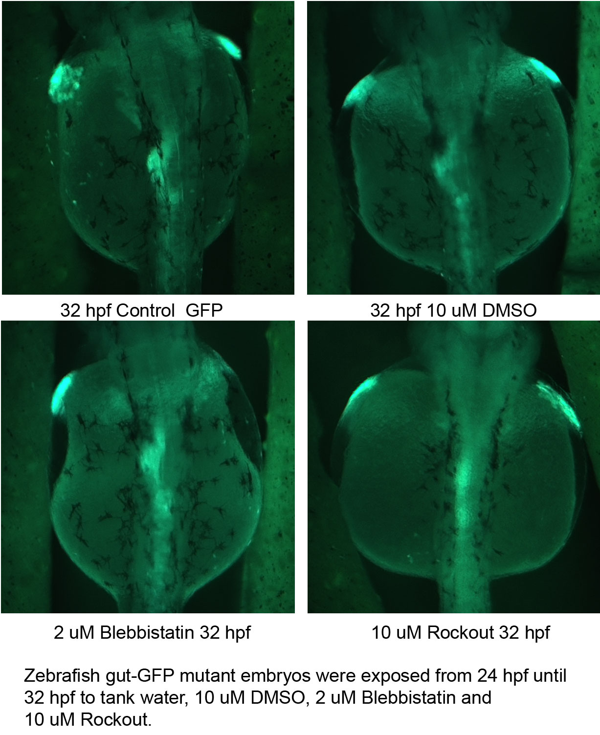
I also had a transgenic zebrafish line that has been modified so that its early gut tube expresses green fluorescent protein (GFP) along with the other normal gut proteins. When the embryos are exposed to short-wavelength light, the GFP fluoresces allowing me to visualize the gut tube.
The results from the GFP embryos mirror what I found with the WISH experiments. Once again, this is a top (dorsal) view of 32 hour post-fertilization embryos. The bright green along the center of the embryo is the gut tube fluorescing. The two control embryos and the Blebbistatin-treated embryos exhibit gut-looping. The Rockout-treated embryos do not.
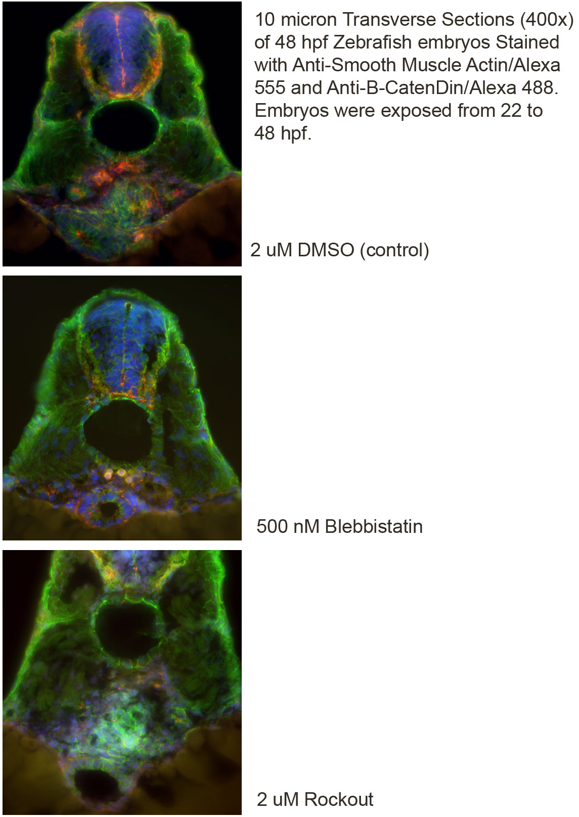
I also sectioned embryos and did immunohistochemistry (IHC) on the embryos to determine the effect of the chemical genetic compounds on gut morphology. Below are a few of the first images I took of the embryos:
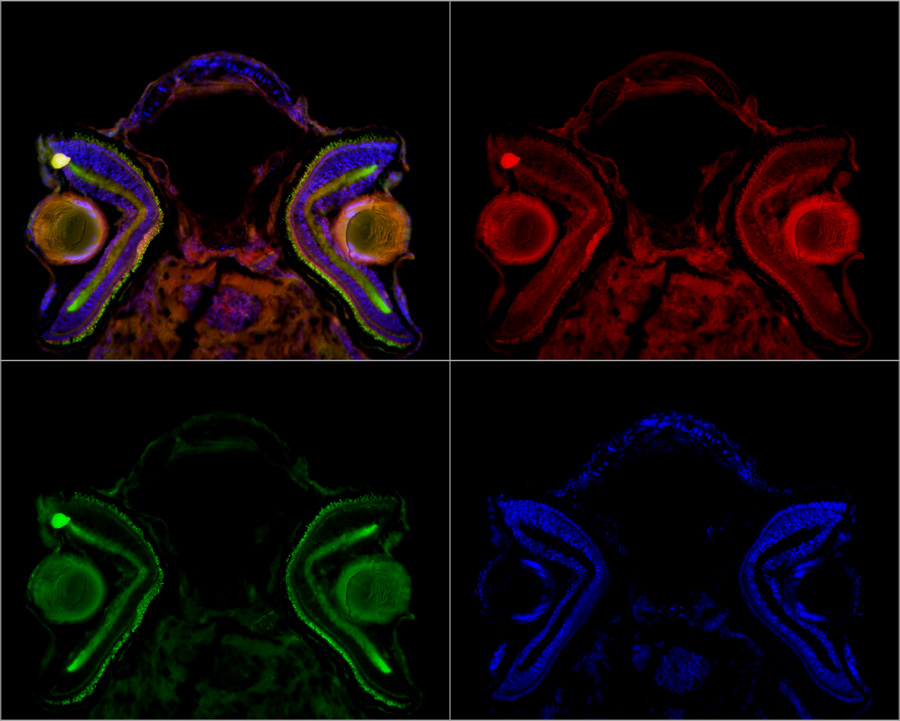
The image below is a composite section through the head and eyes of a 5-day post-fertilization zebrafish larvae. The slide has been anti-smooth muscle actin (red), anti-B-catenin (green) and DAPI which stains the DNA in the cell nucleus. To get the image in the upper left, you have to photograph the section using three different wavelengths of fluorescent light, then combine the images. I was not working on the head region in this project, but from previous work I know that retinoic acid can modulate lens development in Xenopus, so it was interesting to see the lens in the eyecup of these stained sections.
|